Update on Digital Imaging.
Digital Retinal Imaging
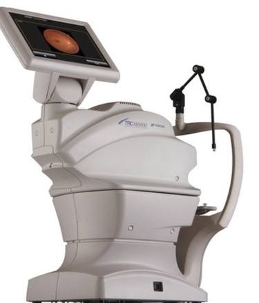
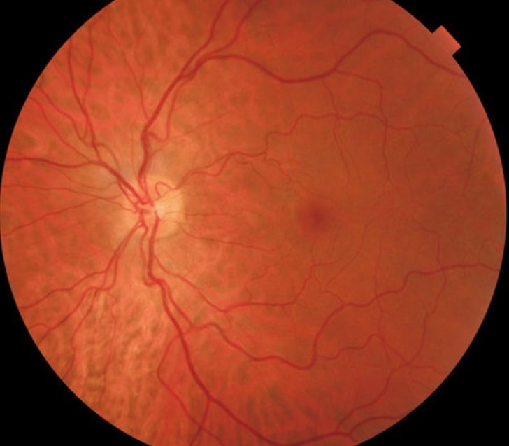
Most of you already know that we have stored retinal images digitally for over twelve years. Our server now holds the retinal details of over thirty thousand patient visits over this period. Our film records are even older and we still use these from time to time. We are now in the process of installing the latest upgrade to our camera systems,

This will enable even more images to be taken without the use of drops to enlarge the pupils. A further significant upgrade will be the option to obtain stereo images to help with the detection of small changes to the optic disc. This should allow us to monitor greater detail early glaucomatous changes.

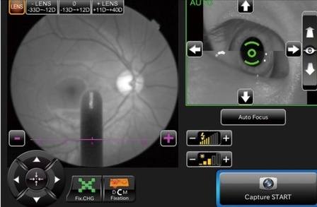
The vast majority of procedures involve no more than a short flash of light to capture the image. This is totally pain free and has only the briefest after image effect, similar to having a portrait photograph taken.

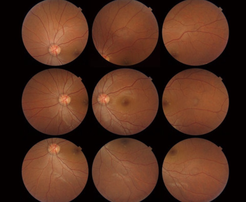
Another very useful function is to extend the normal 45 degree field of view to obtain a much wider view of the peripheral fundus.

The eye ages just like the rest of the body and changes gradually throughout life. These images allow us to record at each visit exactly what is happening to the eyes.